Hyperemia vs. Congestion: The Core Differences
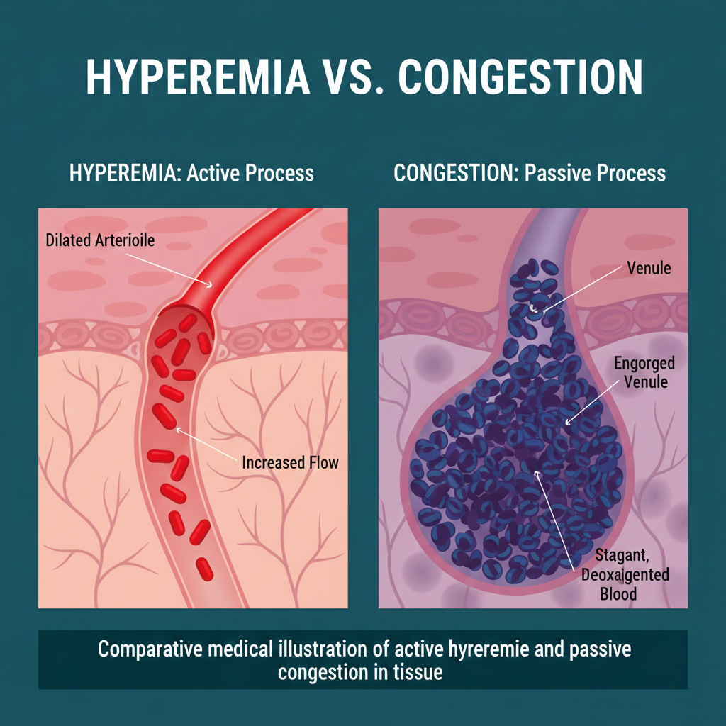
Though both terms describe an organ with “too much blood,” the process behind them is what sets them apart.
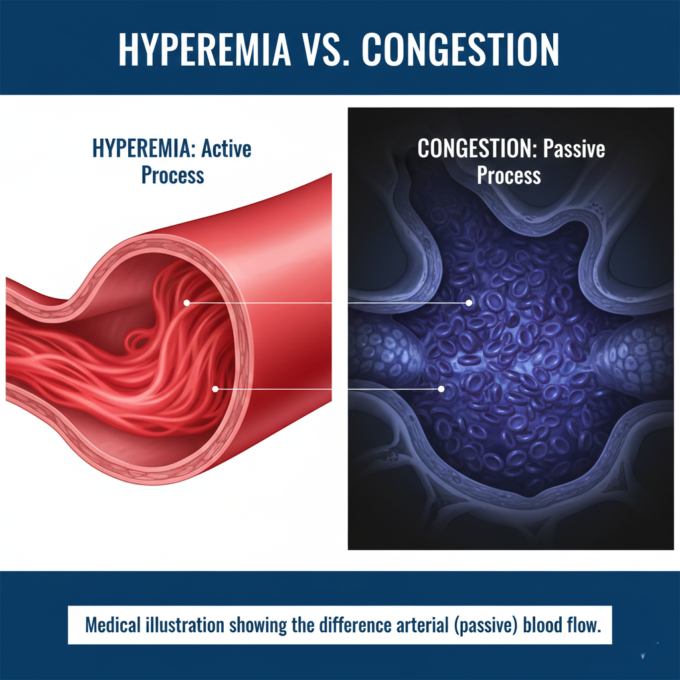
- Hyperemia: This is an active process. It occurs due to arteriolar dilation, which increases blood flow into the tissues. Because this involves oxygenated “pure” blood, the affected organ appears bright red (erythema). Common examples include skeletal muscle during exercise or the redness of a menopausal flush.
- Congestion: This is a passive process. It results from impaired outflow of venous blood from a tissue. This blood is “impure” or deoxygenated, giving the organ a blue-red or dusky appearance known as cyanosis. This is often systemic, caused by heart failure, or localized, caused by a venous obstruction.

Chronic Venous Congestion (CVC)
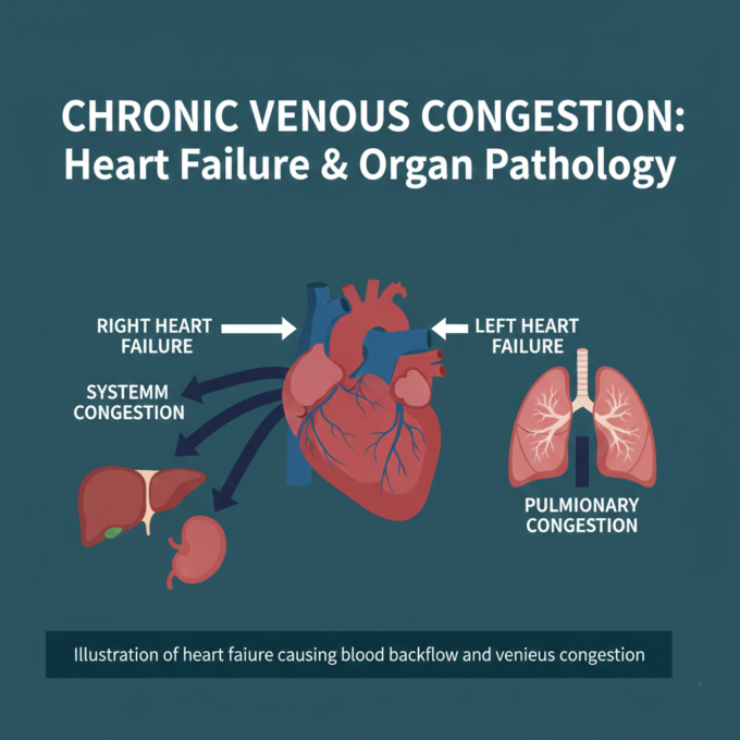
Chronic Venous Congestion (CVC), also known as chronic passive congestion, is a major clinical topic because it reflects how a failing heart affects other vital organs.
CVC of the Lung (Brown Induration)
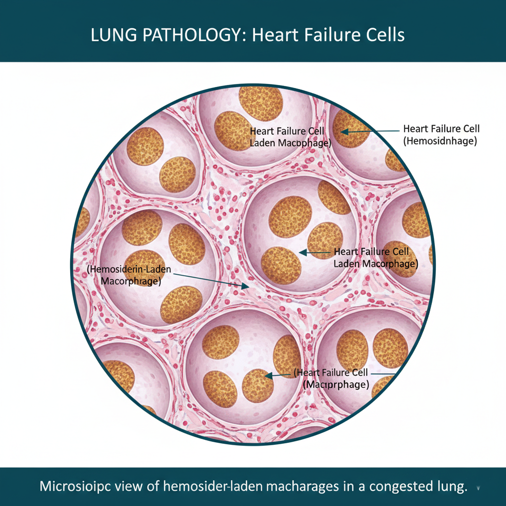
Typically caused by left-sided heart failure, CVC of the lung involves blood backing up into the pulmonary system.
- Gross Appearance: The lungs become heavy, firm, and brownish in color.
- Microscopy: The hallmark of this condition is the presence of “Heart Failure Cells.” When pulmonary capillaries rupture due to high pressure, red blood cells escape into the air sacs (alveoli). Macrophages then ingest the resulting hemoglobin, becoming laden with golden-brown hemosiderin.
CVC of the Liver (Nutmeg Liver)
Caused primarily by right-sided heart failure, blood backs up into the inferior vena cava and eventually the hepatic veins.
- The Nutmeg Appearance: The liver develops an alternate red-and-yellow pattern.
- Mechanisms: The “red” areas are the centrilobular zones (near the central vein) that undergo hemorrhagic necrosis due to deoxygenated blood pooling. The “yellow” areas are the peripheral hepatocytes that undergo fatty change because they are farther from the immediate congestion but still stressed.
CVC of the Spleen
Also a result of right-sided heart failure or portal hypertension, the spleen becomes enlarged (splenomegaly) and firm.
- Gamma-Gandi Bodies: A unique feature in chronic splenic congestion is the formation of Gamma-Gandi bodies. These are small, brown-yellow fosi consisting of old hemorrhage, fibrous tissue, and calcium deposits.
- Microscopy: You will observe a significantly enlarged red pulp (congested sinusoids) compared to the white pulp.
Clinical Summary
Understanding whether a patient’s symptoms are due to an active response (Hyperemia) or a passive failure (Congestion) is the first step in diagnosing systemic issues like cardiac or renal failure. By recognizing the specific patterns—such as heart failure cells in the lungs or the nutmeg liver—clinicians can accurately trace pathology back to its root cause.


Leave a comment